Jaw Fractures and Oral Tumors: When a Dental Specialist Is Critical for Your Pet
A jaw fracture or an oral tumor is not a routine dental problem. These are complex conditions that affect how your pet eats, drinks, breathes, and experiences daily life, and the treatment approach can make a significant difference in both short-term recovery and long-term quality of life. Jaw fractures can result from trauma, but they also happen when advanced periodontal disease (infection of the gums and bone supporting the teeth) weakens the jawbone to the point where even normal chewing causes a break. Oral tumors, whether benign or malignant, can grow quickly and invade surrounding bone, making early detection and precise surgical planning essential.
At North Bay Veterinary Dentistry, we specialize in oral and maxillofacial surgery for exactly these situations. Our advanced diagnostic imaging, including Cone Beam CT (CBCT, a 3D imaging technology that shows the full structure of the jaw, teeth, and surrounding tissues), allows us to see the complete picture before we develop a treatment plan. If your pet has been diagnosed with a jaw fracture or an oral mass and your primary veterinarian has recommended specialist evaluation, call us at (707) 400-0038 or request an appointment to schedule a consultation.
Understanding Jaw Fractures in Pets
How Jaw Fractures Happen
Mandibular fractures in dogs and cats occur from a range of causes, and not all of them involve dramatic accidents.
Traumatic causes:
- Motor vehicle accidents
- Falls from significant height (particularly in cats)
- Bite wounds during fights
- Blunt force trauma to the face
Disease-related fractures: When bone density has been severely reduced by untreated disease, jaw fractures can occur from normal daily activities like chewing or playing. Advanced periodontal disease is a particularly important and often underappreciated cause. As infection destroys the bone supporting the teeth, the jaw becomes structurally compromised. Small and toy breeds see significantly higher rates of severe bone loss around the lower canine teeth. Oral tumors or bone tumors can also weaken the jaw to the point that a fracture can result from what seems like ordinary chewing.
Dog chews that are excessively hard, including antlers, raw bones, and hard nylon products, can also produce fractures directly, both of the teeth and, in extreme cases, of weakened jaw structures.
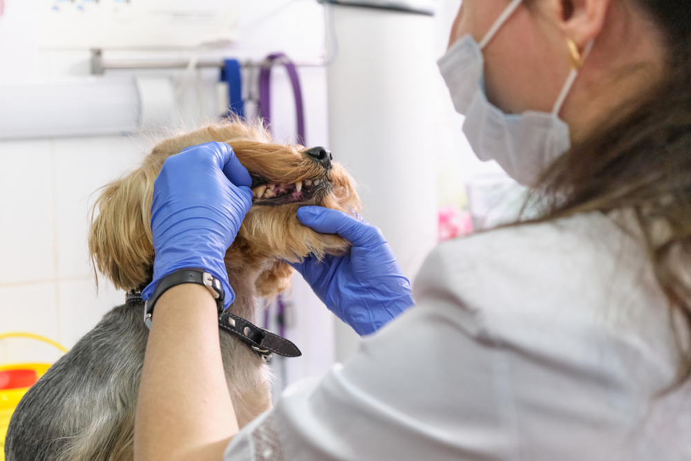
Signs of a Jaw Fracture
Jaw fractures produce signs that range from obvious to surprisingly subtle, particularly when pets are masking pain.
Obvious signs:
- Visible misalignment of the jaw or teeth that do not meet normally
- Inability to close the mouth fully or properly
- Abrupt increase in drooling
- Bleeding from the mouth
- Swelling or bruising around the face or chin
Subtle signs that also warrant prompt evaluation:
- Reluctance to eat, eating more slowly, or dropping food
- Pawing at the face or mouth
- Preference for soft food or avoiding toys
Do not delay evaluation if any of these signs are present. Some fractures that appear manageable from the outside involve significant bone displacement or instability that accelerates damage with every jaw movement. Contact us at (707) 400-0038 if you’re worried.
What Are Oral Tumors, and How Do They Present?
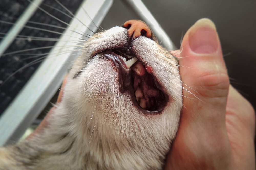
Oral tumors can develop anywhere in the mouth, including the gums, tongue, hard palate, soft palate, and tonsils. Some grow visibly outward from the gum surface while others expand inward, invading bone or soft tissue well before they become apparent on visual examination. Many pets show few obvious signs until a tumor has reached significant size, which is one of the most compelling arguments for routine professional dental evaluation under anesthesia.
Signs that warrant oral evaluation without delay:
- A visible growth, lump, or discolored tissue patch on the gums, tongue, or inside the cheek
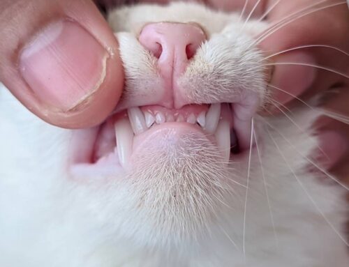
- Loose teeth without obvious periodontal disease to explain them
- Persistent or worsening bad breath that dental cleaning has not resolved
- Difficulty chewing, dropping food, or eating only on one side
- Blood-tinged saliva or unexplained drooling
- Swelling or asymmetry affecting the face or jaw
- Reluctance to allow mouth examination or pawing at the face
Even benign masses can cause pain, displacement of teeth, and secondary infection. “Benign” describes the tumor’s behavior, not the effect on the patient’s comfort or function.
Types of Oral Tumors: Dogs and Cats Differ Significantly
The tumor types seen in dogs and cats are distinct enough that they deserve separate discussion. Species, age, breed, and location all influence the clinical picture, prognosis, and surgical approach.
Common Oral Tumors in Dogs
Dogs develop several primary oral malignancies, and their behavior varies considerably by type. Tumor location and size both strongly influence surgical options and expected outcomes.
- Squamous cell carcinoma in dogs: the second most common oral malignancy in dogs; locally invasive with a tendency toward bone involvement; tonsillar SCC carries a significantly worse prognosis than rostral jaw SCC due to early metastatic spread
- Melanoma: the most common malignant oral tumor in dogs overall; highly aggressive with frequent early metastasis to regional lymph nodes and lungs; requires staging before surgical planning, including thoracic imaging and lymph node assessment
- Fibrosarcoma: locally invasive soft tissue tumor that consistently appears less aggressive than it behaves; surface appearance routinely underestimates depth of invasion, which is why imaging prior to surgery is non-negotiable
- Acanthomatous ameloblastoma: locally aggressive but non-metastatic; derived from dental epithelium; tends to invade bone extensively while remaining histologically benign; wide surgical excision produces excellent long-term results
- Odontogenic fibromas: benign but capable of causing significant tooth displacement and bone remodeling; treatment is surgical with generally favorable prognosis
Large-breed dogs have higher rates of fibrosarcoma. Cocker Spaniels, Weimaraners, and several other breeds have documented increased incidence of melanoma and epulides.
Common Oral Tumors in Cats
Oral cancers in cats are particularly challenging because cats are skilled at concealing discomfort. Reduced grooming, quieter behavior, and subtle changes in eating habits are often the first signs, long before obvious oral pathology is visible. By the time clinical signs are unmistakable, significant disease is often already present.
- Squamous cell carcinoma in cats: accounts for the majority of feline oral malignancies; highly locally invasive with early bone involvement; carries a guarded prognosis even with aggressive treatment; associated with secondhand smoke exposure in some studies; location under the tongue (sublingual) makes early visual detection particularly difficult
- Fibrosarcoma in cats: less common than SCC but similarly aggressive; requires wide surgical margins; can recur locally even after apparently complete excision
The prognosis for feline oral SCC is one of the most sobering in veterinary oncology, which makes early detection through regular professional examination all the more critical.
Why Advanced Diagnostics Are Essential
What Dental Radiographs and Imaging Reveal
Surface examination provides a fraction of the clinical picture. Dental radiographs show what lies beneath the visible surface: the extent of bone loss around a fracture, tumor invasion into alveolar bone, the precise location and margins of a lesion, and the health of surrounding teeth that may need to be addressed as part of treatment.
For fracture repair, knowing exactly where the fracture lines are and how much viable bone remains determines which repair technique is appropriate. For tumor surgery, confirming bone involvement before the procedure determines whether resection must include bone and how wide the margins must be. Surgical planning without complete diagnostic imaging is estimation. Surgical planning with complete imaging is precision.
When CT Imaging Changes the Surgical Plan
CT scans provide three-dimensional imaging that standard radiographs cannot replicate. They are indicated when:
- A fracture involves complex anatomy or multiple segments
- A tumor is large, deeply positioned, or suspected of bone invasion
- Lymph node involvement needs to be assessed before surgery
- Surgical reconstruction of the jaw or palate is planned
- Relationships between the lesion and adjacent critical structures need to be understood before incisions are made
Our CBCT technology produces detailed three-dimensional reconstructions of the jaw and oral structures, providing the information needed to plan complex procedures with precision.
What Does the Diagnostic Workup Involve?
A complete diagnostic workup for an oral mass typically includes sedated oral examination, dental radiographs or CBCT imaging, fine needle aspiration or incisional biopsy for histopathology, and bloodwork. Staging for malignant tumors adds thoracic radiographs and regional lymph node assessment.
It is worth noting that not every oral mass is a tumor, and some things that don’t look like a tumor can be cancerous. Oral inflammatory and ulcerative diseases, like stomatitis, can produce lesions that closely mimic neoplasia on visual examination. Histopathology is the only reliable way to distinguish between them, which is why biopsy is a standard part of evaluation for any new or changing oral mass rather than an optional step.
Jaw Fracture Repair Techniques
The repair approach depends on fracture location, complexity, bone quality, and the patient’s overall health.
Common techniques:
- Interdental wiring combined with acrylic splints for additional stability
- Bone plating with small titanium plates and screws for complex or unstable fractures
- External fixators placed through the bone connected externally when internal fixation is not appropriate
- Acrylic splints bonded to teeth for immobilization, particularly useful in cats and small dogs with limited bone volume
Every case involves individual factors. Fractures complicated by severe periodontal disease, missing teeth, or compromised bone density require technique adaptation that a general approach cannot accommodate.
Oral Tumor Surgery: Margins, Resection, and Realistic Outcomes
Surgical treatment of malignant oral tumors requires wide margins, meaning removal of not just the visible tumor but a substantial surrounding margin of normal tissue to reduce the risk of local recurrence. When tumors have invaded bone, this means mandibulectomy (removal of part of the lower jaw) or maxillectomy (removal of part of the upper jaw).
These procedures sound alarming, but the functional and quality-of-life outcomes are consistently better than many families anticipate. Quality-of-life studies show that dogs who undergo partial jaw resection for cancer report similar scores to unaffected dogs once healed, particularly when clean margins are achieved. The alternative, inadequate margins or delayed surgery, produces worse outcomes in every measure.
Reconstructive options exist for some cases to restore function and appearance following resection. Radiation therapy and chemotherapy may be recommended alongside surgery depending on tumor type, staging results, and proximity to critical structures.
Anesthesia and Pre-Surgical Planning
Our anesthesia protocols are tailored to each patient’s specific health profile, including pre-anesthetic evaluation, IV fluid support, continuous vital sign monitoring, and dedicated anesthesia monitoring throughout the procedure. Nerve blocks are used for regional pain control, reducing the total amount of systemic analgesia required and improving recovery comfort significantly.
Pre-surgical consultation includes review of all diagnostic imaging, discussion of the planned procedure, realistic outcome expectations, and time for questions. Our surgery day expectations page walks families through the day so there are no surprises.
Recovery After Oral Tumor Surgery
What to Expect at Home
Feeding during recovery: most patients require a soft or liquid diet for several weeks. For patients whose oral pain prevents adequate voluntary intake, E-tubes (esophagostomy feeding tubes placed in the neck) provide reliable, comfortable nutritional support without requiring the patient to swallow. E-tube management is straightforward and can be taught to most families before discharge.
Pain management: multimodal pain protocols are used during and after surgery. Most patients are comfortable quickly after surgery, and the goal is maintaining that comfort consistently through the healing period.
Activity restriction: jumping, running, and contact with other pets should be limited during the healing period to protect the repair.
Monitoring the surgical site: redness, swelling, discharge, and any loose wires or implants are reasons to contact us before the scheduled recheck rather than waiting.
Scheduled rechecks evaluate healing, review pathology results if pending, and assess comfort. For tumor cases, ongoing monitoring includes periodic oral examination and imaging to detect local recurrence early, when intervention remains most effective.
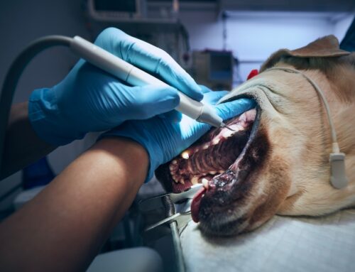
What Routine Dental Care Prevents
Many of the cases we see, including pathological fractures and tumors identified only after significant growth, share a common thread: they were not caught early because routine professional dental examination was not occurring. A comprehensive dental cleaning with full-mouth radiography performed on a regular schedule finds early-stage masses, bone loss patterns that predict fracture risk, and lesions that cannot be seen without imaging or without the level of oral access that anesthesia provides.
The goal of routine preventive care is not just clean teeth. It is consistent surveillance of the entire oral cavity under conditions that allow complete examination. That is how most early-stage oral pathology is actually found.
Frequently Asked Questions
Can my general practice veterinarian handle a jaw fracture or oral tumor?
General practitioners handle the initial assessment, diagnosis, and stabilization. Complex fracture repair and tumor resection are specialist procedures that benefit from training in oral and maxillofacial surgery, specialist-level equipment, and advanced imaging. Your primary veterinarian’s referral is the appropriate pathway when these cases exceed general practice scope.
How do I know if my pet’s mass needs biopsy?
Every new or changing oral mass should be evaluated and biopsied. Clinical appearance alone cannot reliably distinguish benign from malignant. Biopsy determines tissue type and drives the entire treatment plan.
What if my pet has other health problems?
Pre-anesthetic evaluation and tailored protocols address underlying health conditions. Many older or medically complex patients undergo successful oral surgery when approached with appropriate preparation and monitoring. The greater risk in most patients is the untreated pathology rather than the anesthesia required to address it.
What does a consultation involve?
A consultation at North Bay Veterinary Dentistry includes examination, review of prior imaging and records from your primary veterinarian, discussion of findings, and a treatment recommendation with realistic outcome expectations. Our team takes time to ensure you understand the options and what recovery involves before any decisions are made.
Specialist Care When It Matters Most
Jaw fractures and oral tumors are the situations where specialist training, advanced imaging, and surgical precision change outcomes most significantly. At North Bay Veterinary Dentistry, we provide the comprehensive evaluation and precise surgical care these cases require, working in close coordination with your primary veterinarian throughout.
Contact us at (707) 400-0038 or request a consultation to discuss your pet’s diagnosis and options.

Leave A Comment