Resorptive lesions, also known as feline odontoclastic resorptive lesions (FORLs), are a common and painful dental condition affecting cats. These lesions cause progressive destruction of the tooth, leading to pain, inflammation, and potentially severe oral health issues. As a pet owner or a referring veterinarian, understanding resorptive lesions is crucial for early diagnosis and treatment, which can significantly improve a cat’s quality of life.
What Are Feline Resorptive Lesions?
Feline resorptive lesions are characterized by the breakdown of the tooth’s structure, starting at the enamel and progressing into the dentin and root. This process is primarily driven by the activity of odontoclasts, cells that resorb or break down the mineralized tissues of the teeth. The exact cause of this process remains unclear, but it is believed to involve a combination of genetic, environmental, and dietary factors.
Signs and Symptoms of Resorptive Lesions in Cats
Detecting resorptive lesions can be challenging since many cats may not show obvious signs of pain. However, some common indicators include:
- Difficulty Eating: Cats may chew on only one side of the mouth or drop food while eating.
- Decreased Appetite: Pain from eating can lead to reduced food intake.
- Excessive Drooling: In more advanced disease, oral ulcers can cause pain and subsequent drooling.
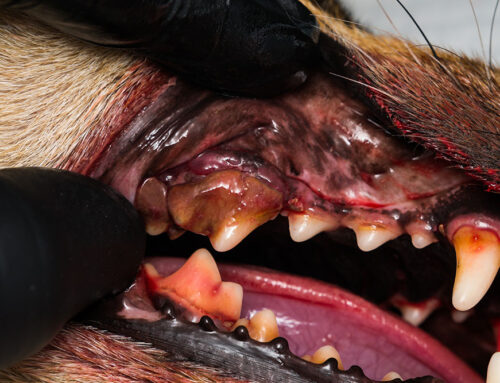
- Visible Red Lesions or Swelling: On the gums, particularly around the affected teeth.
- Tooth Fracture or Loss: A tooth may break suddenly, or you may notice missing teeth during a routine examination.
Diagnosis of Resorptive Lesions
A thorough oral examination under anesthesia, coupled with dental imaging, is essential for diagnosing resorptive lesions. Digital imaging plays a critical role in identifying lesions that are not visible to the naked eye. At North Bay Veterinary Dentistry, we utilize advanced diagnostic tools to detect and assess the severity of these lesions, ensuring the most effective treatment plan.
Treatment Options for Resorptive Lesions
The treatment of feline resorptive lesions typically involves either extraction or crown amputation, depending on the type and extent of the lesions.
- Extraction: This is the most common and definitive treatment for resorptive lesions. Complete extraction of the affected tooth, including the root, is performed to eliminate pain and prevent further complications.
- Crown Amputation with Intentional Root Retention: In cases where the root is ankylosed (fused to the bone), crown amputation may be performed, leaving the root in place to avoid excessive trauma. This approach is often chosen when the root is extensively resorbed, and extraction could cause more harm than benefit.
Post-Treatment Care and Management
After treatment, regular follow-up visits and home care are essential to ensure healing and prevent future lesions. Key aspects of post-treatment care include:
- Regular Check-Ups: Regular dental check-ups help monitor healing and detect any recurrence early.
- Home Oral Hygiene: Although cats can be challenging to brush, using dental gels or feeding them a veterinary-recommended dental diet can help reduce plaque and tartar accumulation. For more tips, visit our guide on brushing your pet’s teeth.
- Monitor for Signs of Pain or Discomfort: Keep an eye out for any signs of oral pain, such as pawing at the mouth or reluctance to eat.
Preventing Resorptive Lesions in Cats
While there is no guaranteed way to prevent resorptive lesions, several strategies may help reduce the risk:
- Regular Monitoring: Frequent vet visits to monitor your cat’s dental health, especially if they are predisposed to dental issues, can lead to early detection and more effective treatment.
- Routine Dental Care: Professional cleanings as recommended, and at-home oral hygiene can help manage oral health throughout your pet’s life.
- Dietary Management: Feeding a diet formulated to support dental health may help slow the progression of dental diseases. Consider incorporating dental chews or toys that encourage chewing and reduce plaque buildup.
Why Choose North Bay Veterinary Dentistry for Your Cat’s Dental Care?

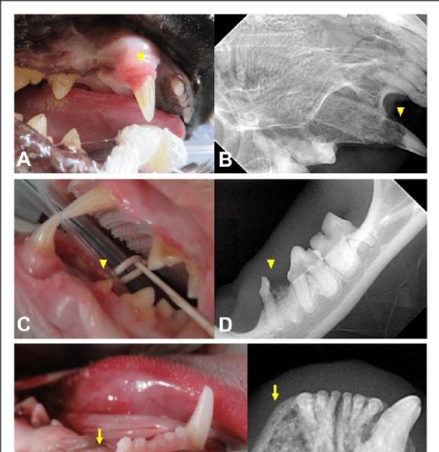
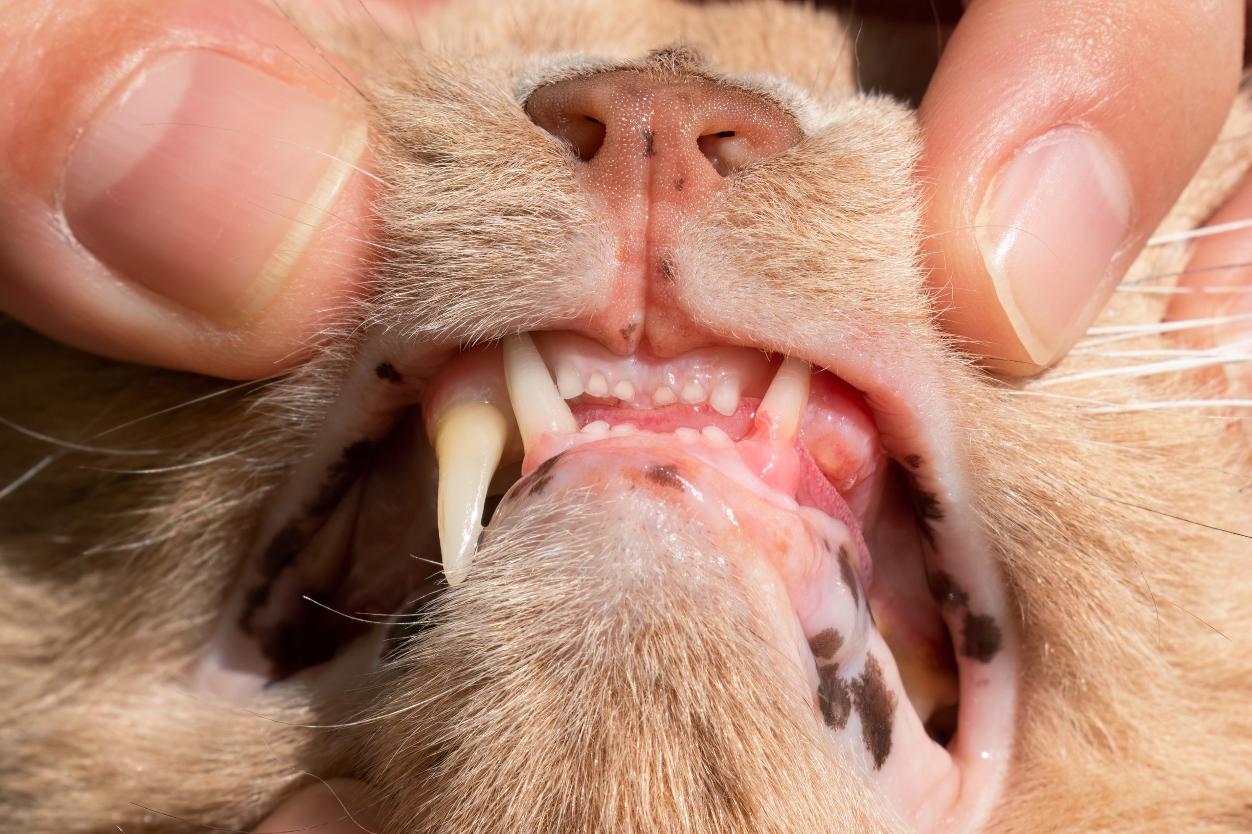
Tooth resorption. A/B) Clinical photograph and radiograph of maxillary right canine tooth (104) showing stage 4c, type 2 TR (arrowheads). C/D) Clinical photograph and radiograph of missing crown of mandibular left third premolar tooth (307) showing stage 4a, type 3 TR (arrowheads). E) Clinical photograph of missing mandibular right canine tooth (404) (arrow). F) Radiograph showing missing 404 with osteitis and stage 4c, type 1 TR of image E (arrow). ————— -Please make “north bay 2_2 imagery” the main photo, no text for the juvenile perio. The other image will go in the article, please use the other text as a caption. text for “imagery for juvenile perio…” Juvenile periodontitis in a 1-year-old male neutered Maine Coon. Note the severe gingivitis affecting the entire attached gingiva, areas of gingival recession and gingival enlargement (Perry R, Tutt C. Periodontal disease in cats: Back to basics – with an eye on the future. Journal of Feline Medicine and Surgery. 2015;17(1):45-65.)
At North Bay Veterinary Dentistry, located in Petaluma, California, we specialize in comprehensive dental care for cats and dogs. Our team includes board-certified veterinary dentists who are experienced in diagnosing and treating feline resorptive lesions and other complex dental conditions. We use state-of-the-art technology, including digital imaging and anesthesia protocols tailored to each patient’s needs, to ensure the highest level of care.
For more information about our services or to schedule a consultation, visit our appointment request page or contact us directly.
Conclusion
Resorptive lesions in cats are a painful and common dental condition that requires early diagnosis and appropriate treatment to prevent significant health issues. By understanding the signs, causes, and treatment options, pet owners and veterinarians can work together to provide the best possible care for cats suffering from this condition. If you suspect your cat may have a resorptive lesion, don’t hesitate to reach out to our team at North Bay Veterinary Dentistry.
Image attribution: Images included in this blog are from the American Veterinary Dental College and “Dental Pain in Cats: A Prospective 6-Month Study” Palmeira, Fonseca, Maria & Lecuelle, (2022). Dental Pain in Cats: A Prospective 6-Month Study. Journal of Veterinary Dentistry.


Leave A Comment