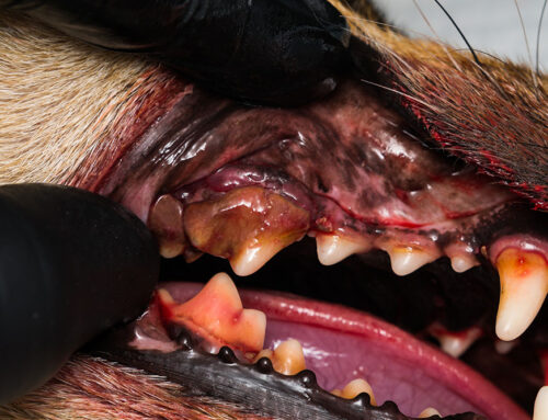
You likely know that veterinary dentists can treat pets’ advanced or complicated periodontal diseases, but you may not be familiar with these specialists’ full skill set or experience. At North Bay Veterinary Dentistry, our veterinary dentist not only treats pets with routine oral issues but also offers specialized imaging and treatment for traumatic facial and jaw fractures.
What is maxillofacial trauma in pets?
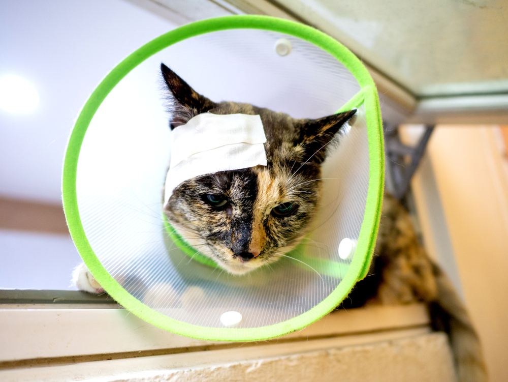
Maxillofacial trauma is the term that describes injuries to a pet’s head, face, and jaw. The most common dog and cat maxillofacial trauma causes include animal bites, falls, and vehicle collisions. Facial injuries can be obvious, such as a displaced jawbone, or they may have a more subtle appearance. Most pets who have suffered a trauma have multiple injuries to several body areas but these aren’t always apparent. Seeking veterinary care immediately after an accident is vital to rule out facial injuries as well as life-threatening internal organ or brain trauma.
Diagnostic imaging for maxillofacial trauma in pets
Diagnostic imaging for maxillofacial injuries can be tricky. Traditional X-rays can identify some injuries, but the many facial and skull bones overlap in these images, which can hide fractures or make them difficult to localize. If your pet sustains facial trauma and X-rays or a physical exam indicate the presence or possible presence of fractures, a traditional computed tomography (CT) scan may be recommended. However, imaging for small patients or injuries concentrated in the teeth and jaws may require referral to our facility for cone beam CT (CBCT) and intraoral X-rays.
What are intraoral X-rays for pets?
Intraoral X-rays are a veterinary dentistry cornerstone that are similar to the X-rays you get when you go to the dentist. These images provide a two-dimensional view of the teeth, roots, and surrounding soft tissues. Our veterinary dentist places a small plate strategically behind or under each suspected injury area, which prevents the overlapping views a skull X-ray causes and eliminates distortion. Intraoral X-rays allow us to evaluate tooth and tooth root integrity in healthy pets and in those who have sustained facial or oral trauma. However, intraoral X-rays provide a limited view at one time and these images aren’t enough to fully evaluate traumatic injuries. In these cases, our team combines X-rays with CBCT.
What is cone beam computed tomography for pets?
CBCT has become widely available in veterinary medicine and is particularly useful for veterinary dentistry. The technology is similar to traditional CT scanners, which use radiation to take image “slices” and combine the slices to create a three-dimensional image. CBCT does this as well but on a smaller scale. The technology takes image slices that are less than one millimeter, compared with a standard CT’s one- to three-millimeter imaging slices, making CBCT especially useful for imaging pets’ teeth and jaws, especially those who are extremely small.
CBCT images the entire skull and can detect traumatic fractures’ exact location and extent. In many maxillofacial trauma cases, pets have multiple fractures throughout the skull that may or may not require specific treatment or monitoring. We combine CBCT with X-rays to get a detailed view of each tooth to plan surgical dental treatments and fracture repairs. This advanced technology provides high-resolution images at a lower cost, with less radiation exposure, and in less time than other advanced imaging techniques, allowing for more efficient treatment and better outcomes.
Our North Bay Veterinary Dentistry team uses only the most advanced diagnostic equipment and procedures to ensure accurate facial trauma and oral condition diagnosis and treatment. Contact us to learn more about our services or to schedule a consultation with our experienced and compassionate team.


Leave A Comment